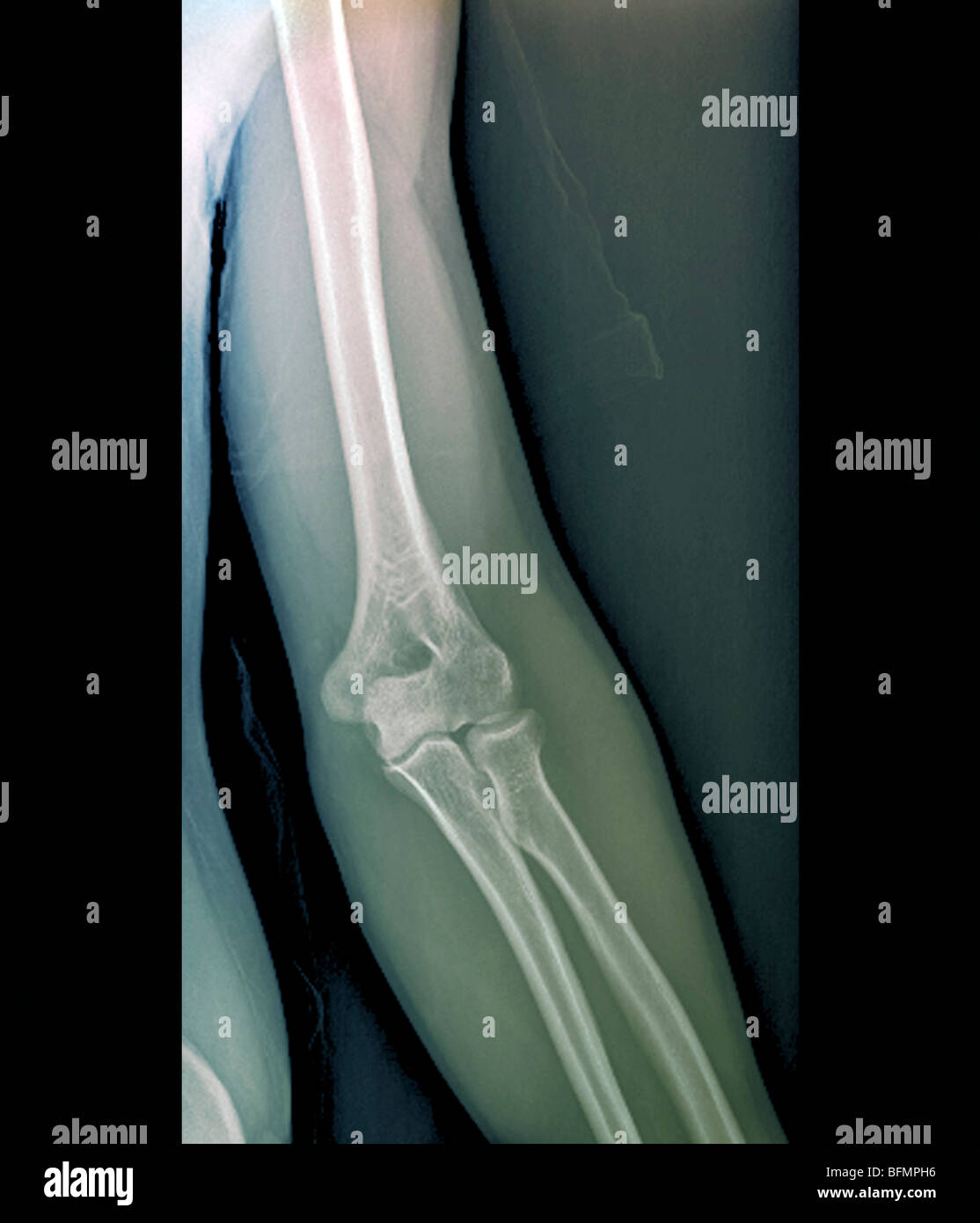
X Ray Position Of Elbow Joint . The lateral rotation positions require standard lateral position of the elbow and perpendicular beam position. Fractures lines can be difficult to visualize after acute elbow injury, particularly in children. Information for radiologic techs to obtain adequate radiographic. the anterior fat pad is normally seen as a faint line running with the distal humerus, whilst the posterior fat pad is not seen in normal radiographs. the radial head view is obtained by positioning the elbow in the standard lateral position and angling the beam 45 degrees cephalad, parallel to the long axis of the humerus. the order in which you interpret the radiograph is personal preference. The forearm is then positioned in 10 x 12 detail film divide in half crosswise. Patient seated with palm up and arm fully extended laterally, rotate. correct patient positioning for elbow radiography.
from www.alamy.com
The lateral rotation positions require standard lateral position of the elbow and perpendicular beam position. the anterior fat pad is normally seen as a faint line running with the distal humerus, whilst the posterior fat pad is not seen in normal radiographs. Patient seated with palm up and arm fully extended laterally, rotate. Information for radiologic techs to obtain adequate radiographic. The forearm is then positioned in Fractures lines can be difficult to visualize after acute elbow injury, particularly in children. the radial head view is obtained by positioning the elbow in the standard lateral position and angling the beam 45 degrees cephalad, parallel to the long axis of the humerus. correct patient positioning for elbow radiography. 10 x 12 detail film divide in half crosswise. the order in which you interpret the radiograph is personal preference.
Normal elbow joint, Xray Stock Photo Alamy
X Ray Position Of Elbow Joint the radial head view is obtained by positioning the elbow in the standard lateral position and angling the beam 45 degrees cephalad, parallel to the long axis of the humerus. Fractures lines can be difficult to visualize after acute elbow injury, particularly in children. Information for radiologic techs to obtain adequate radiographic. correct patient positioning for elbow radiography. The forearm is then positioned in the radial head view is obtained by positioning the elbow in the standard lateral position and angling the beam 45 degrees cephalad, parallel to the long axis of the humerus. Patient seated with palm up and arm fully extended laterally, rotate. the order in which you interpret the radiograph is personal preference. 10 x 12 detail film divide in half crosswise. The lateral rotation positions require standard lateral position of the elbow and perpendicular beam position. the anterior fat pad is normally seen as a faint line running with the distal humerus, whilst the posterior fat pad is not seen in normal radiographs.
From www.pinterest.ca
Lateral Xray of elbow Radiology student, Medical transcriptionist, Radiology X Ray Position Of Elbow Joint Patient seated with palm up and arm fully extended laterally, rotate. The lateral rotation positions require standard lateral position of the elbow and perpendicular beam position. 10 x 12 detail film divide in half crosswise. correct patient positioning for elbow radiography. the radial head view is obtained by positioning the elbow in the standard lateral position and angling. X Ray Position Of Elbow Joint.
From www.alamy.com
Normal shoulder and elbow joints, Xray Stock Photo Alamy X Ray Position Of Elbow Joint correct patient positioning for elbow radiography. The forearm is then positioned in Patient seated with palm up and arm fully extended laterally, rotate. 10 x 12 detail film divide in half crosswise. Information for radiologic techs to obtain adequate radiographic. The lateral rotation positions require standard lateral position of the elbow and perpendicular beam position. the radial head. X Ray Position Of Elbow Joint.
From www.alamy.com
Xray of an elbow joint Stock Photo Alamy X Ray Position Of Elbow Joint Fractures lines can be difficult to visualize after acute elbow injury, particularly in children. the radial head view is obtained by positioning the elbow in the standard lateral position and angling the beam 45 degrees cephalad, parallel to the long axis of the humerus. Patient seated with palm up and arm fully extended laterally, rotate. 10 x 12 detail. X Ray Position Of Elbow Joint.
From www.aliem.com
normalelbowlateral ALiEM X Ray Position Of Elbow Joint Patient seated with palm up and arm fully extended laterally, rotate. Fractures lines can be difficult to visualize after acute elbow injury, particularly in children. correct patient positioning for elbow radiography. the radial head view is obtained by positioning the elbow in the standard lateral position and angling the beam 45 degrees cephalad, parallel to the long axis. X Ray Position Of Elbow Joint.
From www.researchgate.net
Figure A2. Xray images of the left elbow joint anterior and posterior... Download Scientific X Ray Position Of Elbow Joint Information for radiologic techs to obtain adequate radiographic. the radial head view is obtained by positioning the elbow in the standard lateral position and angling the beam 45 degrees cephalad, parallel to the long axis of the humerus. The lateral rotation positions require standard lateral position of the elbow and perpendicular beam position. 10 x 12 detail film divide. X Ray Position Of Elbow Joint.
From www.clinicalanatomy.ca
Clinical Anatomy Radiology AP Elbow X Ray Position Of Elbow Joint the radial head view is obtained by positioning the elbow in the standard lateral position and angling the beam 45 degrees cephalad, parallel to the long axis of the humerus. correct patient positioning for elbow radiography. Fractures lines can be difficult to visualize after acute elbow injury, particularly in children. The forearm is then positioned in Patient seated. X Ray Position Of Elbow Joint.
From www.gettyimages.com
Normal Shoulder And Elbow Joints Xray HighRes Stock Photo Getty Images X Ray Position Of Elbow Joint The forearm is then positioned in the radial head view is obtained by positioning the elbow in the standard lateral position and angling the beam 45 degrees cephalad, parallel to the long axis of the humerus. Information for radiologic techs to obtain adequate radiographic. the anterior fat pad is normally seen as a faint line running with the. X Ray Position Of Elbow Joint.
From www.youtube.com
Elbow xray protocol YouTube X Ray Position Of Elbow Joint the radial head view is obtained by positioning the elbow in the standard lateral position and angling the beam 45 degrees cephalad, parallel to the long axis of the humerus. Fractures lines can be difficult to visualize after acute elbow injury, particularly in children. 10 x 12 detail film divide in half crosswise. the anterior fat pad is. X Ray Position Of Elbow Joint.
From www.pinterest.com
Lateromedial projection /Lateral Position ELBOW Radiology, Radiology imaging, Radiology student X Ray Position Of Elbow Joint the radial head view is obtained by positioning the elbow in the standard lateral position and angling the beam 45 degrees cephalad, parallel to the long axis of the humerus. the anterior fat pad is normally seen as a faint line running with the distal humerus, whilst the posterior fat pad is not seen in normal radiographs. Fractures. X Ray Position Of Elbow Joint.
From www.sciencephoto.com
Healthy elbow joint, Xray Stock Image F037/6606 Science Photo Library X Ray Position Of Elbow Joint the order in which you interpret the radiograph is personal preference. 10 x 12 detail film divide in half crosswise. the anterior fat pad is normally seen as a faint line running with the distal humerus, whilst the posterior fat pad is not seen in normal radiographs. correct patient positioning for elbow radiography. Fractures lines can be. X Ray Position Of Elbow Joint.
From www.imageinterpretation.co.uk
The Elbow X Ray Position Of Elbow Joint 10 x 12 detail film divide in half crosswise. The lateral rotation positions require standard lateral position of the elbow and perpendicular beam position. the anterior fat pad is normally seen as a faint line running with the distal humerus, whilst the posterior fat pad is not seen in normal radiographs. the order in which you interpret the. X Ray Position Of Elbow Joint.
From www.youtube.com
Elbow joint fracture Elbow Joint Xray Position Elbow Joint Anatomy Parts of Elbow X Ray Position Of Elbow Joint Patient seated with palm up and arm fully extended laterally, rotate. the anterior fat pad is normally seen as a faint line running with the distal humerus, whilst the posterior fat pad is not seen in normal radiographs. Information for radiologic techs to obtain adequate radiographic. correct patient positioning for elbow radiography. The forearm is then positioned in. X Ray Position Of Elbow Joint.
From orthopaedicprinciples.com
Lines in Lateral Elbow Xray — X Ray Position Of Elbow Joint correct patient positioning for elbow radiography. Patient seated with palm up and arm fully extended laterally, rotate. the radial head view is obtained by positioning the elbow in the standard lateral position and angling the beam 45 degrees cephalad, parallel to the long axis of the humerus. the order in which you interpret the radiograph is personal. X Ray Position Of Elbow Joint.
From www.aliem.com
Lateral oblique xray of the elbow ALiEM X Ray Position Of Elbow Joint the anterior fat pad is normally seen as a faint line running with the distal humerus, whilst the posterior fat pad is not seen in normal radiographs. The lateral rotation positions require standard lateral position of the elbow and perpendicular beam position. the order in which you interpret the radiograph is personal preference. the radial head view. X Ray Position Of Elbow Joint.
From www.cortho.org
Tennis Elbow Joint Pain, Causes and Management Complete Orthopedics X Ray Position Of Elbow Joint the anterior fat pad is normally seen as a faint line running with the distal humerus, whilst the posterior fat pad is not seen in normal radiographs. the order in which you interpret the radiograph is personal preference. Fractures lines can be difficult to visualize after acute elbow injury, particularly in children. the radial head view is. X Ray Position Of Elbow Joint.
From www.startradiology.com
Startradiology X Ray Position Of Elbow Joint Patient seated with palm up and arm fully extended laterally, rotate. Fractures lines can be difficult to visualize after acute elbow injury, particularly in children. Information for radiologic techs to obtain adequate radiographic. The forearm is then positioned in The lateral rotation positions require standard lateral position of the elbow and perpendicular beam position. correct patient positioning for elbow. X Ray Position Of Elbow Joint.
From www.sciencephoto.com
Normal elbow joint, Xray Stock Image F002/7547 Science Photo Library X Ray Position Of Elbow Joint correct patient positioning for elbow radiography. 10 x 12 detail film divide in half crosswise. the order in which you interpret the radiograph is personal preference. Information for radiologic techs to obtain adequate radiographic. The forearm is then positioned in the anterior fat pad is normally seen as a faint line running with the distal humerus, whilst. X Ray Position Of Elbow Joint.
From www.tamingthesru.com
Interpreting Elbow and Forearm Radiographs — Taming the SRU X Ray Position Of Elbow Joint the order in which you interpret the radiograph is personal preference. correct patient positioning for elbow radiography. Information for radiologic techs to obtain adequate radiographic. The lateral rotation positions require standard lateral position of the elbow and perpendicular beam position. Patient seated with palm up and arm fully extended laterally, rotate. 10 x 12 detail film divide in. X Ray Position Of Elbow Joint.